Dipstick analysis

Another dipstick is applied to the urine (as your GP will have done in the surgery) to check for signs of infection or inflammation (white and/or red blood cells, protein and nitrates) including the pH of the urine. Additionally, the dipstick reagent pads test for the presence of Bilirubin, Urobilinogen, Ketones and Glucose.

Urine pH

A neutral pH is 7.0. The higher the number, the more alkaline it is. The lower the number, the more acidic your urine is. The average urine sample tests at about 5.0 as the urine is slightly acidic. This is due to the normal daily acid production in the body to maintain an acid-base balance. Therefore, any abnormalities in the acid-base balance has a direct effect on urinary pH levels.

These levels are particularly useful in the evaluation of stones, crystals or infection. For example, in a patient with a possible kidney stone, the urinary pH level is helpful. The main types of kidney stones are:

  • calcium stones, the most common type of stone
  • struvite stones, usually caused by an infection, like a urine infection
  • uric acid stones, usually caused by a large amount of acid in your urine

Uric acid, cystine, and calcium oxalate stones tend to form in acidic urine, whereas struvite (magnesium ammonium phosphate) and calcium phosphate stones form in alkaline urine.

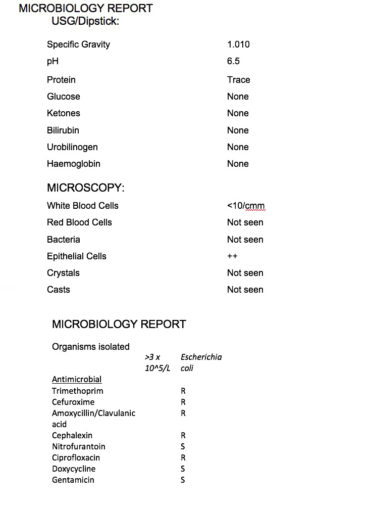
People with a UTI due to Proteus and Klebsiella bacteria typically have alkaline urine whereas bacteria such as E-coli are usually found in a more acidic environment.

This pH will also be noted on the report. However, pH is also affected by diet; a high protein intake can give rise to acidic urine, whereas a high intake of dairy products or vegetables can give rise to alkaline urine.

White Blood Cells (Leukocytes)

Only a few white blood cells are normally present in urine. When these numbers increase, the dipstick test will become positive. This indicates that there is inflammation in the urinary tract or kidneys and the body is excreting more white blood cells. In addition to possible infection, white blood cells/leukocytes can also indicate chronic kidney inflammation caused by a kidney stone, a tumour of the kidneys, bladder or urethra, infections such as chlamydia or other sexually transmitted diseases and fungal infections such as Thrush.

Protein

Protein in the urine may be a sign of kidney disease. The protein test pad provides a rough estimate of the amount of albumin in the urine. Albumin makes up about 60% of the total protein in the blood. Normally, there will be no protein or a small amount of protein in the urine. When urine protein is elevated, a person has a condition called proteinuria.

Small amounts of albumin may be found in the urine when kidney dysfunction begins to develop. If it is felt necessary the laboratory may request that a further, more detailed urine albumin test be carried out by the GP or hospital. The urine albumin test is more sensitive than a dipstick urinalysis and is routinely used to screen people with chronic conditions that put them at risk for kidney disease, such as diabetes and high blood pressure. Protein in the urine can also be due to dehydration, pregnancy, disease of the heart and some cancers.

Nitrates

Urine will contain a certain amount of nitrate usually down to foodstuffs but, in a normal urine sample, this type of nitrite will be indicated as ‘absent’ or ‘not present’ on a dipstick. But, in certain instances, nitrates can be indicated as being ‘present’ or “+”. The most common occurrence of positive nitrites in urine is in the presence of bacteria which convert the non-ionic nitrate into nitrite.

Bacteria where nitrates would be shown as positive on dipstick include certain species of E. coli, Klebsiella, Proteus or Pseudomonas.

However not all bacteria convert nitrates in the urine so relying on this marker in the testing process does not exclude infection.

An important point to note is the conversion of nitrates in the urine can take up to four hours and a fresh sample of urine may not readily show this if the sample is immediately analysed by the laboratory.

Bilirubin and urobilinogen

Bilirubin is a chemical produced when red blood cells are broken down. It is transported in the blood to the liver, where it is processed and excreted into the gut as a constituent of bile. In the gut, bacteria act on the bilirubin to transform it into urobilinogen. It is usual for urine to contain urobilinogen but not bilirubin. Bilirubin in the urine may be an indicator of a breakdown of red blood cells. It may not be effectively removed by the liver, which may suggest liver disease or a problem with drainage of bile into the gut, such as gall stones.

Ketones

These are chemicals that are formed during the abnormal breakdown of fat and are not normal constituents of urine.

Breakdown of fat may result from prolonged vomiting, fasting or starvation; individuals on a diet or who present with diarrhoea and vomiting may have a positive result.

Ketones can also be present in the urine of people with poorly controlled diabetes. This can make the blood more acidic and is known as diabetic ketoacidosis; it should be reviewed urgently by a doctor. Some medications, such as captopril, may also produce a false positive result (Steggall, 2007).

Glucose

Glucose in the urine (glycosuria) can occur in pregnancy or patients taking corticosteroids. It may also be indicative of diabetes but is not normally found in a urine sample. Although glycosuria is an indication of endocrine abnormality, it is not diagnostic and further investigation, such as fasting blood tests, may be required.

Urine microscopy

Once this dipstick check has been carried out, a microscopic examination is the next step using urine sediment. To do this, the urine sample is centrifuged (spun) to concentrate the substances in it at the bottom of a tube. The fluid at the top of the tube is then disposed of and the drops of fluid remaining are examined under a microscope and the following will be noted on your urine sample report:

Red blood cells (RBCs)

Normally, a few RBCs are present in urine. Inflammation, injury, or disease in the kidneys or elsewhere in the urinary tract, for example in the bladder or urethra, can cause RBCs to leak out of the blood vessels into the urine. However, RBCs can also be due to blood from haemorrhoids or menstruation, kidney, bladder or urethral cancers, enlargement of the prostrate in men; kidney stones and certain blood diseases such as sickle cell anaemia.

White blood cells (WBCs or leukocytes)

The number of WBCs in urine is normally low. When the number is high, it indicates an infection or inflammation somewhere in the urinary tract due to the immune system response.The agreed total number of leukocytes to be present in a millilitre of urine to establish the baseline for infection remains unchanged since the work of C Dukes in 1928. He proposed a threshold of less than 10 wbc/per millilitre of urine as the upper limit of normal white blood cell excretion in the urine. Above that limit and the sample may be positive for infection.

Also note that White Blood Cell or Leukocytes can also indicate chronic kidney inflammation caused by a kidney stone, a tumour of the kidneys, bladder or urethra, infections such as chlamydia or other sexually transmitted diseases and fungal infections such as Thrush.

Epithelial cells

Epithelial cells can be either squamous, originating from the vagina, urethra or genital skin, urothelial, that is cells from the bladder wall and finally renal tubular originating from the kidneys.When analysing the sample, significant epithelial cells may lead to further analysis to identify their type. Squamous epithelial cells originate from the urethra or vagina and are commonly found in urine samples due to transfer whilst providing the sample – these are often reported as a contaminate in the sample without specific analysis of the actual type of epithelial cell. However, it’s the presence of urothelial epithelial cells that would indicate a urinary tract infection. Or, if many renal (kidney) epithelial cells are discovered in urinalysis, then it could indicate a viral infection or problem with the kidneys.

Casts

Casts are particles that are formed from protein secreted by kidney cells. Under the microscope, they often look like a sausage shape because of the way they form and in healthy people they appear nearly clear. This type of cast is called a ‘hyaline’ cast. When an infection is present in the kidney, other things such as RBCs or WBCs can become trapped in the protein as the cast is formed. When this happens, the cast is identified by the substances inside it, for example as a red blood cell cast or white blood cell cast. Different types of casts are associated with different kidney diseases and the type of casts found in the urine may give clues as to which is affecting the kidney. Normally, healthy people may have a few casts. After strenuous exercise, more casts may be detected. Cellular casts, such as RBC and WBC casts, indicate a kidney disorder.

Crystals

Urine contains many dissolved substances (solutes) – waste chemicals that your body needs to eliminate after filtration through the kidneys.These can form crystals and they may group together to form kidney “stones”. These stones can become lodged in the kidney itself or in the ureters – tubes that pass the urine from kidney to the bladder – causing extreme pain. Medications, drugs, and x-ray dye can also crystallize in urine.

Bacterial incubation on petri dish

If the microscopic examination shows signs of infection then a sample of the urine sediment will be placed on a Petri dish and left to incubate over 18 -24 hours. Bacteria can reproduce very quickly given the right conditions, such as warmth, moisture and suitable nutrients.

To ensure the cultures are not contaminated by other microorganisms, the following sterile conditions are needed; the Petri dishes, nutrient agar jelly inside these dishes and other culture media must be sterilised.

The inoculating loops used to transfer microorganisms to the petri dish must be sterilised (usually by passing the metal loop through a Bunsen burner flame) and finally, the lid of the Petri dish is sealed with sticky tape to stop microorganisms from the air getting in and contaminating the culture.

Bacteria will produce colonies differing in appearance as they grow, some colonies may be coloured, some colonies are circular in shape, and others are irregular. Different bacterial strains produce these characteristics. The sample from the petri dish is then “gram stained” to identify the actual strain of bacteria. This means a crystal violet colour stain is applied to the sample and placed under a microscope.

All bacteria are described as either Gram-negative or Gram-positive

Gram-positive bacteria remain purple. Gram-negative bacteria are stained pink. Gram-negative bacterial strains include:

  • Escherichia coli
  • Klebsiella pneumoniae
  • Proteus mirabilis
  • Pseudomonas species
  • Morganella morganii
  • Citrobacter species

Gram-positive strains include:

  • Enterococcus faecalis
  • Enterococcus faecium (E. faecium)
  • Staphylococcus aureus
  • Staph saprophyticus

Under the microscope, the appearance of bacteria is observed. The lab can then finally determine:

  • Are they Gram-positive or negative?
  • What are the physical characteristics?
  • Are the cells individual or are they in chains, pairs etc.?
  • How many are there and how large are the cells?

The final part of the process is to then count the total colony numbers of the single bacteria identified.

These are known as colony-forming units usually abbreviated as CFU. For a positive diagnosis to be made of an infection, the agreed standard is currently greater than >10 5 /ml in a millilitre of urine. Up to 10,000 colonies of bacteria/ml are considered normal. Greater than 100,000 colonies/ml represents a positive urinary tract or kidney infection.

For counts between 10,000 and 100,000, the culture is indeterminate and results will show low growth or mixed growth.

If sufficient bacteria are grown and the single pure strain identified e.g. E-coli, antibiotics will be tested against the bacteria to check their effectiveness in stopping the infection. This is known as susceptibility and resistance and the results will be noted on the lab report to help your GP prescribe the correct antibiotic.

Yeast can also be present in urine. If yeast is found in urine, then the laboratory may recommend tests for a yeast (fungal) infection on vaginal secretions or may culture on a petri dish to identify the yeast and whether it could be causing UTI or other disease symptoms.

Antibiotic testing/bacterial susceptability

If sufficient bacteria are grown and the single pure strain identified such as e coli or enterococcus, antibiotics will be tested against the bacteria to check their effectiveness in stopping the infection. This is known as susceptibility and resistance and the results will be noted on the lab report to help your GP prescribe the correct antibiotic.

To determine bacterial susceptibility, the laboratory injects the bacteria isolated from the sample into a series of tubes or cups that contain broth dilutions of the antibiotic. After a standardized incubation period, the lowest concentration of antibiotic that prevents visible growth of the organism is classified as the minimal inhibitory concentration (MIC).

The alternate method of bacterial susceptibility is via the disk diffusion method. Using this technique, disk plates are impregnated with various antibiotics and placed on the surface of an agar plate that has been injected with the isolated bacterial sample. The antibiotic diffuses outward from the disk over a standard incubation time, and the diameter of the zone of inhibition is measured. The size of this zone is compared with standards to determine the sensitivity of the organism to the drug.

The laboratory report that will be sent to your GP will report the name of bacterium grown and the sensitivities of the antibiotics tested against each bacterium.

The interpretation of this testing categorizes each antibiotic result as susceptible (S), intermediate (I), sensitive-dose dependent (SD), resistant (R) or no interpretation (NI).  What does this mean?

  • Susceptible (S): This indicates that the antibiotic may be an appropriate choice for treating the infection caused by the bacteria tested. i.e. the organism is likely to respond to treatment with this drug, at the recommended dosage. Bacterial resistance is absent or at a clinically insignificant level.
  • Intermediate (I): It is applicable to those is bacteria that are “moderately susceptible” to an antibiotic. The intermediate category serves as a buffer zone between susceptible and resistant. The antimicrobial agent may still be effective against the tested isolate but response rates may be lower than for susceptible isolates.
  • Susceptible-dose dependent (SDD):
    This is a new category for antibacterial susceptibility testing. If a particular bacteria falls under this category, the susceptibility will depend on the dosing regimen used. Higher doses or more frequent doses or both should be used to achieve concentration levels that are more likely to be clinically effective.
  • Resistant (R). If a bacteria is resistant to a particular antibiotic; it won’t be inhibited by that specific medication using a normal dosage. There is also no expectation of this bacteria to respond a higher dosage.
  • Non-susceptible (NS) This category is used for bacteria where there are resistant strains.


Microbiology report for GP and patient

Once the laboratory analysis has been finished the report is sent to your GP. If the report is positive for bacterial infection, you will be contacted to visit the GP and a prescription issued for the identified antibiotic from the laboratory report to treat your UTI. If the report details the possibility of kidney stones, you may be referred by the GP for further tests and treatment particularly if the stone is causing considerable pain. If the sample is negative, then if symptoms persist, you may request a retest from your GP.

A sample report:

sample urine microbiology report

Expanded Quantitative Urine Culture (EQUC)

Increasingly used in research studies investigating the urinary microbiome and how it influences recurrent and chronic UTI, Expanded Quantitative Urine Culture (EQUC) is a new technology using a used a modified culture protocol from that of the standard urine analysis laboratory method.  This includes analysing larger volumes of urine (10–100 mL instead of 1 mL for a standard mid stream urine culture), the incubation of samples using differing atmospheric conditions to cultivate bacteria that may not grow on standard urine culture and longer times for incubation where the urine sample is incubated in a special liquid broth.  If no growth is seen after 3 days, the incubation is extended for another 3 days. For slower growing bacteria or those where different methods of development other than standard agar plate are needed, EQUC is a useful tool.

A study by Loyola University Chicago, published in the American Journal of Microbiology in 2014 comparing the standard urine culture against EQUC noted that the EQUC analysis method found bacteria in 92% of samples tested compared against the same samples analysed via standard methods which showed negative growth for bacteria. With this, bacterial presence as low as 10 colony forming units (CFU) per mL could be detected; indeed, Lactobacillus, Corynebacterium, and multiple other genera were isolated using this EQUC. The authors proposed a “streamlined” version of this EQUC, which specified using a higher volume of 100 mL of urine on MacConkey, blood, and colisitin-nalidixic acid (CNA) agars in a 5% CO2 incubator for 48 h to yield 84% sensitivity relative to the extended spectrum protocol. This protocol has been utilized by several investigators since.

A further study of 150 women published in the Journal of Clinical Microbiology in 2016, patients were grouped by whether or not they had self-reported UTI-like symptoms and performed the aforementioned streamlined EQUC on catheterized urine specimens; while they did not find a difference in the number of isolated uropathogens, they did find a reduced species richness and diversity in patients who did have clinical UTI symptoms. About half of the uropathogens in the UTI cohort were missed by standard urine culture; additionally, the threshold of 105 CFU/mL would not report a predominant organism in numerous patients with a clinical UTI in this cohort.

PCR or polymerase chain reaction testing

PCR testing (polymerase chain reaction) is a technique used to amplify trace amounts of DNA found in or on almost any liquid or surface.

Every human, animal, plant, parasite, bacteria or virus contains genetic material such as DNA sequences that are unique to their species.

If a urine sample contains segments of DNA, PCR is a method used to amplify (make many more identical copies) of these unique sequences to determine with a very high probability the pathogenic bacteria, fungi or other microbes.

Within urine, PCR testing has detected the DNA of more than 1,200 microbial species.

Benefits of PCR testing

  • PCR tests are not affected by temperature or time delays unlike traditional urine cultures where the urine sample must be delivered to the laboratory within two hours to prevent sample degradation.
  • The results of a PCR test can be available within 24 hours.
  • PCR tests do not grow microbes, they are instead extracting the microbial DNA from a sample and can identify multiple bacteria, anaerobes or fungi.
  • A PRC test needs one sample unlike standard laboratory tests where a separate swab would be required for fungal microbe analysis in addition to a urine sample.  This fungal analysis can take up to 20 days to process.

Limitations of PCR testing

  • Laboratories using this method need to preselect which microorganisms the test looks for.  Urine panels usually comprise of less than a dozen micro-organisms, more often the most common for that type of bacterial infection. Any other organisms not listed in this panel will be excluded from analysis.  It amounts to around 1% of all known microbes.
  • It is currently unknown how the urinary microbiome may differ from the microbiome of the bladder wall and in particular those bacteria embedded into bladder wall cells or those affected by biofilm coverage. To understand the differences further will likely require more invasive sampling procedures such as biopsies.
  • The DNA extraction technique chosen will significantly affect how faithfully the bacterial composition of the original sample is represented by the DNA extracted from it. In some bacteria, such as Gram-positive bacteria and Mycobacteria, it can be more difficult to break down the cell membrane of the bacteria (known as lysis) for study than others in a microbial community and these bacteria may be less represented in a report. On the other hand, if an extraction method is too harsh, the DNA from the easily lysed species may become sheared. Currently, there is no standard technique that works equally well for lysing all bacteria in a given sample.
  • Another limitation of sequencing-based approaches is that they only yield information about the microbial DNA in a sample. While this allows for identification of the types of bacteria present in a biological sample, it does not distinguish between bacteria that are live from those that are dead.
  • In comparison to Next Generation Sequencing testing it is thus limited because it won’t offer a genetic report of everything in the urine sample you provide.
  • PCR reports give percentages on the bacteria or microbes found in your urine. But the understanding of the urinary microbiome is still in development. Are all those identified responsible for your urine infection? Does a higher percentage equate to the causative agent behind your infection?
  • The testing methods are based on individual, unique, patented methods used by each laboratory. They test against a panel of microbes/fungi/anaerobes established by that company. If the infection causing agent is not found against that panel, how are you to be treated
  • There is also the issue of cost. Treatment of a chronic UTI may involve several tests over a period of time and at present, these are extremely expensive. Some patients in the US are able to offset their costs through health insurance but others are unable to do so. International patients cannot offset against health insurance.
  • It can be difficult to find a practitioner who can interpret the laboratory findings and treat accordingly. Treatment guidelines used by GPs and consultants worldwide are based on standard testing protocols. Some patients have found their results have been dismissed or ignored by their doctor or consultant.
  • Finally, as yet, no research studies have been published using PCR testing in a clinical setting to determine successful treatment outcomes for patients using this diagnostic method.

Next Generation Sequencing (NGS)

Bacteria can be classified using conventional microbiology methods, such as microscopy or being grown on specific media in a laboratory and using antibiotic sensitivity assessments. In recent decades, molecular microbiology methods have revolutionized bacterial identification. A popular method is 16S ribosomal RNA (rRNA) gene sequencing. This method is not only faster and more accurate than conventional methods, but also allows identification of strains that are difficult to grow in standard laboratory conditions.

The 16S rRNA gene is present in all bacteria. This makes it an ideal genetic fragment to be used in identification and comparison of bacteria. NGS is a process which uses this 16S rRNA gene identification to analyse for quality. Only the highest quality bacteria, anaerobes or fungi identified will be interpreted and reported. The data for each detected bacterial or fungal species is then reported as a percentage of specimens identified.

Deep NGS or Shotgun sequencing goes further, it creates a genetic fingerprint of everything in your urine sample and is able to identify tens of thousands of microorganisms in one sample. This can include not only bacteria but parasites and viruses something that PCR and simple 16S NGS testing cannot offer.

Benefits of NGS testing

  • NGS tests are not affected by temperature or time delays unlike traditional urine cultures where the urine sample must be delivered to the laboratory within two hours to prevent sample degradation. Often standard cultures will return a no growth result when samples have not arrived at the laboratory within the time frame required for successful culturing.
  • NGS testing does not grow microbes, they are instead extracting the microbial DNA from a sample and can identify multiple bacteria, anaerobes or fungi. They require just need one sample.
  • These reports will be comprehensive in nature covering all micro-organisms in your sample rather than focusing on a single pathogen which the standard laboratory culture is directed towards.

Issues with NGS testing

There are some uncertainties about NGS testing in a clinical setting:

  • All testing is reliant on the sample containing the presence of biofilm pieces or infected bladder lining cells.  If these are not present in the sample you submit, no laboratory test offered by these companies will be able to detect the pathogens contained within them. Dormant, embedded bacterial DNA will not be detected.
  • At present, it isn’t possible for a DNA sequencing test to tell you whether any micro-organisms identified were part of a biofilm or intracellular community or not.
  • It is currently unknown how the urinary microbiome may differ from the microbiome of the bladder wall and in particular those bacteria embedded into bladder wall cells or those affected by biofilm coverage. To understand the differences further will likely require more invasive sampling procedures such as biopsies.
  • The DNA extraction technique chosen will significantly affect how faithfully the bacterial composition of the original sample is represented by the DNA extracted from it. In some bacteria, such as Gram-positive bacteria and Mycobacteria, it can be more difficult to break down the cell membrane of the bacteria (known as lysis) for study than others in a microbial community and these bacteria may be less represented in a report. On the other hand, if an extraction method is too harsh, the DNA from the easily lysed species may become sheared. Currently, there is no standard technique that works equally well for lysing all bacteria in a given sample.
  • Another limitation of sequencing-based approaches is that they only yield information about the microbial DNA in a sample. While this allows for identification of the types of bacteria present in a biological sample, it does not distinguish between bacteria that are live from those that are dead.
  • NGS reports give percentages on the bacteria/fungi or microbes found in your urine. But the understanding of the urinary microbiome is still in development. Are all those identified responsible for your urine infection? Does a higher percentage equate to the causative agent behind your infection?
  • The testing methods are based on individual, unique, patented methods used by each laboratory.  They test against a panel of microbes/fungi/anaerobes established by that company.  If the infection causing micro-organism is not found against that panel, how are you to be treated?
  • There is also the issue of cost. Treatment of a chronic UTI may involve several NGS tests over a period of time and at present, these are extremely expensive. Some patients in the US are able to offset their costs through health insurance but others are unable to do so. International patients cannot offset against health insurance.
  • It can be difficult to find a practitioner who can interpret the laboratory findings and treat accordingly. Treatment guidelines used by GPs and consultants worldwide are based on standard testing protocols. Some patients have found their results have been dismissed or ignored by their doctor or consultant.
  • Finally, as yet, no research studies have been published using NGS testing in a clinical setting to determine success outcomes for patients using this diagnostic method.

Fresh urine microscopy

A sample of fresh urine is dropped onto a plate for immediate analysis under a microscope. White blood cells and epithelial cells are counted as markers of an infection. When infection occurs in the urinary tract, the immune system tries to remove infected cells by shedding the cells in the bladder lining (the epithelium) to be excreted during urination – these are known as epithelial cells.

A high white blood cell count usually indicates that the body is fighting an infection. White blood cells rush in to help destroy the harmful substance and prevent the infection developing further.

White blood cells degrade very quickly. Studies have shown this occurs in as little as four hours unless stored in the correct conditions so a sample sent for analysis at the laboratory will not pick them up.

Professor James Malone-Lee, Emeritus Professor of Nephrology, UCL notes in two published studies: “Analysis of urothelial and clue cells is novel, as is sediment culture, but these methods have been well validated and have a strong pathophysiological foundation. Fresh urine microscopy is not commonly adopted in clinical practice nowadays, nevertheless, it has been well validated in studies dating back to 1928 and is still unsurpassed as a surrogate marker of infection”. (1)

“Pyuria, detected by microscopy of a fresh midstream urine (MSU) specimen, is the most sensitive surrogate marker of UTI. It circumvents the problems associated with quantitative (numerical) bacterial culture, and its value in the diagnosis of UTI is recognised by international practice guidelines. Whilst ≥10 wbc in a micro litre of urine is employed almost universally to diagnose UTI, contemporary data cast doubt on this threshold in patients with LUTS. In the symptomatic patient, controlled studies have demonstrated that lower pyuria counts of 1–9 wbc in a micro litre of urine are associated with an increase in independent inflammatory and microbiological markers of UTI. Thus, lower levels of pyuria may also indicate infection and immune activation”.(2)

Issues with urine microscopy

  • Urine samples must be fresh, that is within two hours of urination to prevent degradation of the white blood cells otherwise the sample must be discarded
  • Training of staff to interpret samples via microscopy plus the cost of the relevant microscope
  • Acceptance by clinicians of the results and then appropriate treatment prescription. At present urine testing is biased towards bacterial identification. Urine microscopy, once the standard for urine analysis, was superseded by the automated methods introduced since the 1960s preferred for their time-saving benefits.

References:

1. A blinded observational cohort study of the microbiological ecology associated with pyuria and overactive bladder symptoms. Kiren Gill, Ryoon Kang, Sanchutha Sathiananthamoorthy, Rajvinder Khasriya, James Malone-Lee. International Urogynecology Journal, January 2018

2. Recalcitrant chronic bladder pain and recurrent cystitis but negative urinalysis: What should we do?  Sheela Swamy, William Barcella, Maria De Iorio, Kiren Gill, Rajvinder Khasriya, Anthony S. Kupelian,  Jennifer L. Rohn, James Malone-Lee. International Urogynecology Journal, March 2018

Phage therapy treatment – would it work for UTI?

There is a lack of peer-reviewed controlled clinical trials making it difficult to properly evaluate the effectiveness of such therapeutics by western standards; more observational studies of phage therapy in patient treatment are needed for it to be considered safe and effective for usage in the treatment of urinary tract infections. Currently, their use is limited and they are an unlicensed treatment.